ExoFlow-ONE EV Labeling Kit for Flow Cytometry (Emerald Green)
| Specifications | |
|---|---|
| Product Category: | Exosome Labeling for Flow Cytometry |
Product Description
- Directly detect EVs by specifically labeling internal EV components
- Accurately estimate the size of labeled EVs
- Ensure proper run-to-run calibration with the included size standards
- Learn more from a single sample by multiplexing with our range of spectrally separated ExoFlow-ONE dyes
- Achieve near single-vesicle visualization* with our proprietary ExoFlow-ONE dyes that deliver
> High quantum efficiency
> Negligible background signal when not bound to EVs
Finally, you can take full advantage of the power of flow-based methods for analyzing extracellular vesicles (EVs) with SBI’s exclusive ExoFlow-ONE Gemstone dyes. By specifically labeling internal EV components with one of these proprietary, high quantum efficiency dyes, you can achieve near single-vesicle resolution* for more powerful flow cytometry and FACS studies, and uncover greater insights into EV biology.
The ExoFlow-ONE Emerald Green Gemstone Dye Kit comes with enough labeling reagents for 25 reactions**. Each kit also includes 1.5 mL of silica bead size standards (110 nm – 1300 nm), two of which are fluorescent. The silica beads, sourced from Apogee Flow Systems, are specifically designed to produce a refractive index and light scattering patterns similar to biological particles, enabling more accurate estimation of EV particle size.
*Requires a specialized flow cytometry imager capable of high-resolution vesicle analysis (e.g. Amnis® ImageStreamX Mark II, Apogee Micro-PLUS).
**Each reaction is defined as 1 µL of working dye combined with 200 – 500 µg of EV protein resuspended in 500 µL of buffer.
Choose the right ExoFlow-ONE dye for your project
| CAT.# | EXOFLOW ONE DYE | EXCITATION/EMISSION WAVELENGTH | RECOMMENDED LASER LINE |
| EXOF100A-1-SBI | Ruby Red | 573/588 nm | 561 nm |
| EXOF200A-1-SBI | Garnet Far Red | 628/643 nm | 633 nm |
| EXOF300A-1-SBI | Emerald Green | 511/525 nm | 488 nm |
| EXOF400A-1-SBI | Sapphire Blue | 403/454 nm | 405 nm |
| EXOF500A-1-SBI | Citrine Yellow | 542/556 nm | 532 nm |
Supporting Data
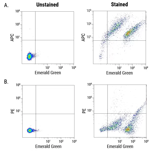
Figure 1. ExoFlow-ONE dyes multiplexed with antibody-conjugated dyes are able to identify subpopulations of EVs. Murine-derived EVs were isolated and co-stained with Emerald Green ExoFlow-ONE and an APC-conjugated antibody against a cell adhesion protein (A) or Emerald Green ExoFlow-ONE and a PE-conjugated antibody against a macrophage membrane protein (B). In panel A, right, two distinct populations of EVs can be seen (Emerald Green axis), each of which expresses a range of the cell adhesion protein (APC axis). In contrast, in panel B, right, while the two distinct populations of EVs can still be seen (Emerald Green axis) the EVs do not appear to express the macrophage-specific protein. Thus, ExoFlow-ONE dyes multiplexed with antibody-conjugated dyes can be used to easily detect subpopulations of EVs.
- Catalog Number
EXOF300A-1-SBI - Supplier
SBI System Biosciences - Size
- Shipping
Blue Ice