Minute Detergent- and EDTA-free Total and Plasma Membrane Protein Isolation and Fractionation Kit (Native Proteins obtained)
| Specifications | |
|---|---|
| Product Category: | Cell Fractionation |
| Sample Type: | Cells, tissues |
Product Description
Invent Biotechnologies Minute™ plasma membrane protein isolation kit is composed of optimized buffers (detergent and EDTA free) and protein extraction filter cartridges with 2.0 ml collection tubes. The kit is designed to rapidly isolate native total membrane proteins (organelle membrane proteins) and native plasma membrane proteins from cultured mammalian cells or tissues.
Due to the use of protein extraction filter cartridges, the membrane protein isolation is simple, easy and user friendly with high yield. Unlike many commercial membrane preparation kits that require large amount of starting cells (5 millions and up) this kit offers a wide range of starting cells (1-50 Million cells/sample). The buffers are detergent and EDTA free. A Dounce homogenizer or a tissue blender is not needed. The procedure can be completed in less than 45 min.

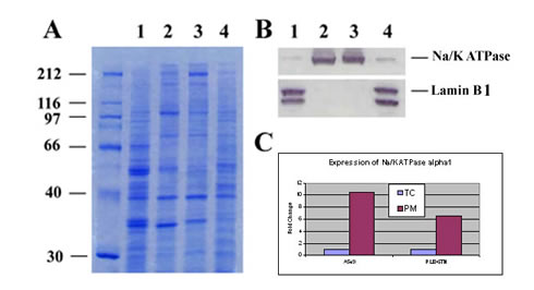
Figure 1:
A. SDS-PAGE profiles of total cell lysate (TC) and isolated plasma membrane proteins (PM) from human and rat cultured cells. Lane 1, Human lung cancer cell A549 total cell lysate; Lane 2, Isolated PM of A549; Lane 3, PM of rat REL-6TN cells; Lane 4, Total cell lysate of rat REL-6TN cells.
B. Western blottings: Proteins in A were transferred to nitrocellulose membrane and probed with anti-Na/K ATPase alpha1, a plasma membrane marker (Upstate, Clone 464.6)) and anti-lamin B1, a nuclear envelope marker (ab16048, abcam Cambridge, MA). The specific protein bands were visualized by a color metric substrate Opti-4CN (Bio-RAD).
C. Densitometry measurement of Na/K ATPase alpha1 signals in B (TC vs. PM). Total cell lysates were extracted by Minute Denaturing Total Protein Extraction Kit (SD-001-IB, Invent Biotechnologies, Eden Prairie, MN).

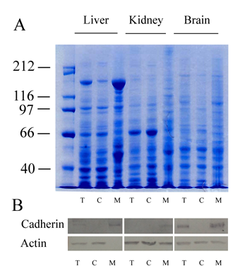
Figure 2:
A. SDS-PAGE profiles of isolated total membrane proteins from mouse tissues (T=Total Cell lysate, C=Cytosol Fraction, M= Total Membrane Fraction)
B. Proteins shown in A were transferred to a nitrocellulose membrane and probed with rabbit-anti mouse pan-cadherin (ab6529,abcam, Cambridge, MA), and anti-actin by Western blotting. The specific protein bands were visualized by a color metric substrate Opti-4CN (Bio-RAD). Signals of pan-cadherin (a 130 kda plasma membrane marker) were significantly enhanced in total membrane protein fractions. Total cell lysates were extracted by Minute™ Denaturing Total Protein Extraction Kit (SD-001-IB, Invent Biotechnologies, Eden Prairie, MN).

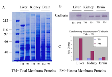
Figure 3:
A. SDS-PAGE profiles of total membrane protein fraction (TM) and isolated plasma membrane protein fraction from mouse tissues.
B. Western blotting of proteins in A were transferred to nitrocellulose membrane and probed with rabbit anti-cadherin (abcam, Cambridge, MA). The specific protein bands were visualized by a color metric substrate Opti-4CN (Bio-RAD).
C. Densitometry measurement of cadherin signals in B (TM vs. PM).
References
- Kutluay, S.B. et al. (2014) Global Changes in the RNA Binding Specificity of HIV-1 Gag Regulate Virion Genesis. Cell 159: 1–14.
- Wen, D. et al. (2013) Regulation of BK-a expression in the distal nephron byaldosterone and urine pH. Am J Physiol Renal Physiol 305: F463-F476.
- Catalog Number
SM-005-IB - Supplier
Invent Biotechnologies - Size
- Shipping
RT

