Basic Fluorescent Protein Vectors
Fluorescent Protein Vectors
Multicolor protein collection for in vivo labeling applications
Evrogen has developed a wide collection of bright basic fluorescent proteins (FPs) for applications in the field of live-cell assays including labeling of cells, subcellular structures, and proteins, analysis of promoter activity and generation of stably transfected cell lines expressing fluorescent proteins or their fusions.
In addition to ready-to-use subcellular localization vectors, vectors comprising fluorescent protein coding sequence linked with multiple cloning sites allow easy targeting of the reporter in the specific cellular compartment. The targeting signal can be fused to C- or N- end of the fluorescent protein using appropriate expression vector (marked with "-C" or "-N" correspondingly). Unmodified vectors can be used for cytosolic expression of fluorescent markers.
Evrogen basic fluorescent proteins are divided into subgroups according to their properties and recommended applications.
TagFPs
Bright monomeric fluorescent proteins of different colors for protein localization and interaction studies: TagYFP, TagRFP, FusionRed
TagFPs group comprises proteins that are optimized for protein localization / interaction studies and stable expression in long term cultures. They can be also used for other cell labeling applications.
TagFPs demonstrate successful performance in fusions with cellular proteins and can be expressed in various heterological systems. Ranging in color from blue to far-red, they provide unique possibilities for multicolor labeling of subcellular structures.
TurboFPs
Superbright and fast-maturing fluorescent proteins for gene expression analysis, cell and organelle labeling applications: TurboGFP, TurboYFP, TurboFP602, Turbo635
TurboFPs are fluorescent proteins of different colors characterized by superbright fluorescence and superior fast maturation. These proteins are recommended for applications requiring fast appearance of bright fluorescence, including cell and organelle labeling or tracking promoter activity.
Far-red and Near-infrared Fluorescent Proteins
Far-red and near-infrared fluorescent proteins for whole body imaging: Katushka2S, TurboFP650
Deep-tissue imaging using fluorescent proteins allows direct and non-invasive observation of the biological processes inside living organisms. The major difficulty of fluorescent imaging within whole animals is the light absorption by melanin and hemoglobin, as well as light scattering. Both absorption and scattering become less pronounced as the light wavelength increases. The optimal "optical window", which is most transparent for the visualization in living tissues, is considered to be between 650-700 and 1100 nm. Therefore, use of far-red or near infrared fluorescent proteins drastically increases sensitivity of the whole body imaging comparing to conventional FPs.
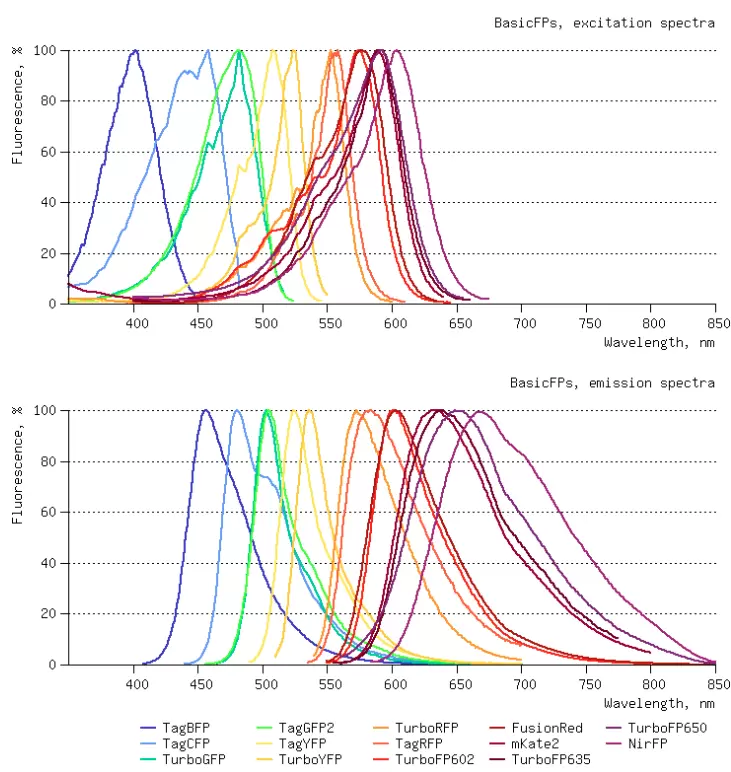
Diversity of Evrogen Basic Fluorescent Proteins
Ranging in color from blue to far-red, Evrogen fluorescent proteins can be used for multicolor labeling to observe different cellular events in a particular cell or a cell population.
