Ready-to-use Subcellular Localization Vectors
Subcellular Localization Vectors
Tools for fluorescence labeling of subcellular structures
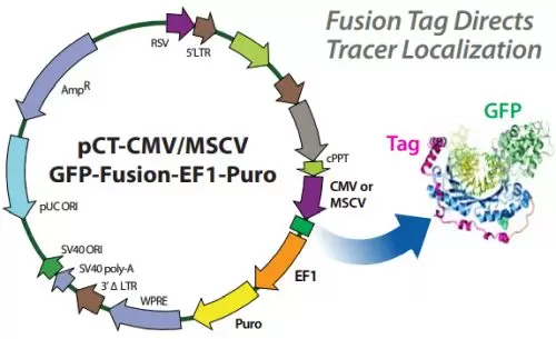
Subcellular localization vectors express a fluorescent protein fused to a specific protein tag directing the fusion protein to the desired subcellular location.
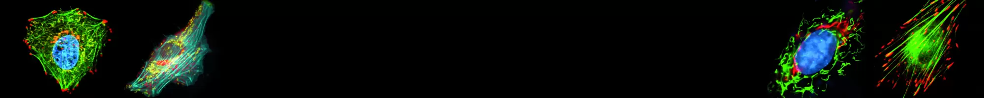
Molecular trafficking is a dynamic process in eukaryotic cells and the Cyto-Tracers provide the ability to light up cell compartments to monitor movement and localization of organelles and to trace endocytosis and exocytosis. Our partner SBI has created a line of lentivector-based Cyto-Tracers that utilize GFP-fusion proteins to mark cellular compartments, organelles, vesicles and structures to enable more long-term and more in-depth experimentation. The Cyto-Tracers can be used in transfections as well as packaged into virus to create stable copGFP or RFP tracer cell lines in primary cells, tumor cell lines and stem cells.
- Stable lentivector-based system
- Great for creating stable reporter cell lines
- Ideal for co-localization studies
- Monitor cellular dynamics and functional studies in real time
- Create your own subcellular label for a broad range of studies
How it works
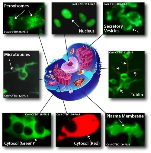
Target Locations
Organelles - Endoplasmic reticulum, endosomes, Golgi apparatus, ER-Golgi vesicles, lysosomes, membrane, mitochondria, peroxisomes, autophagosomes
Exosomal Proteins - CD9 Tetraspanin, CD63 Tetraspanin, CD81 Tetraspanin
Cytoskeletal and adhesion proteins - β-actin, α-tubulin, microtubules
Nuclear Proteins - Histone H2B
Other - Cytosol, plasma membrane, inner leaflet membrane, dendritic membrane
Tracking of subcellular activities
Cyto-Tracers mark your compartment of choice.