SpheroTribe - All-in-one kit for 3D cell culture
Especially useful with challenging cells

Product Description
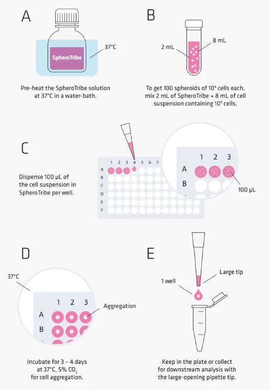
SpheroTribe provides a simple toolkit to generate consistent and robust 3D cell structures. Simply dilute the SpheroTribe solution into your culture medium of choice, watch your cells turn into uniformly sized 3D spheroids and collect them for your downstream assays.
Once diluted in your culture medium of choice, our concentrated polymer-based solution increases the medium viscosity favouring cell-cell contacts. SpheroTribe offers a simple method to generate homogeneous 3D cell structures with increased control over their size and shape, which can be easily handled and washed for downstream experiments.
SpheroTribe is particularly useful to boost aggregation when working with challenging cells, minimize variability between samples and improve the consistency of your migration/invasion assays, immunostaining, drug screening or in vivo implantation experiments.
Kit content
- 25mL of 5X methylcellulose solution
- 10x U-bottom 96-well plates
- 2x racks of 96 pipette tips with a large opening of 200µL
Features
Cell aggregation booster
SpheroTribe provides a gel-like scaffold that favors cell-cell contacts by increasing medium viscosity. It was shown to generate compact spheroids mimicking solid tumors, improve spheroid formation with some of the most challenging cells and to speed up stem cell-derived organoid formation.
Increased homogeneity
By maximizing cell aggregation, SpheroTribe promotes the formation of unique & uniformly sized spheroids allowing for consistent assays (growth, invasion, immune infiltration, in vivo injection, etc).
Easy to use
No need to work on ice, have access to sophisticated equipment of expertise. With SpheroTribe, you have everything on hand to easily grow & handle your spheroids.
Universal
The SpheroTribe solution is composed of methylcellulose, a biologically inert compound dissolved in basal culture medium without any proteins, lipids or growth factors. You can dilute it in any culture medium of your choice, and add additional compounds as desired (i.e. serum, antibiotics, differentiation factors, etc).
How it Works

Cell Type Examples
So far, SpheroTribe has been successfully used for spheroid/organoid formation with the following cell types:
Patient-derived stem-like glioblastoma cells (GB P3 and BL13), human glioblastoma cell lines (U87 & T98G), HeLa, human vaginal mucosal melanoma (HMV-II), human primary colorectal cancer cells, human breast cancer cells (MDA-MB 231), human induced pluripotent stem cells, monkey kidney fibroblast-like cell line (COS-7), primary neurons from rat embryos (E18) & murine melanoma cells (B16F10).
Downstream Experiments
Once spheroids have grown to your desired size, you can use them for any kind of assay according to your regular workflow. The SpheroTribe solution can be readily washed off, leaving a spheroid available for other tests at any stage of your protocol.
Example of in situ assays you can perform directly on the U-bottom plate supplied:
- Live imaging
- Growth/proliferation studies
- Toxicity studies
Examples of downstream assays that might require transferring spheroids to other vessels:
- Invasion & migration assays
- Immunostaining
- Biochemical assays
- Immune infiltration assays
- In vivo implantation
Performance Data

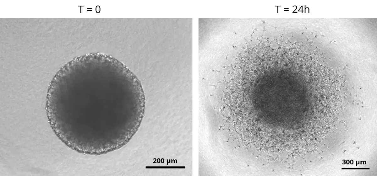
Fig.1. Patient-derived glioblastoma spheroid formed with SpheroTribe invading a 3D collagen-I matrix.
Glioblastoma spheroids were included into a collagen matrix after being cultured in medium added with SpheroTribe solution for 4 days.
Images were taken immediately after (left) or 24 hours after (right) inclusion in the collagen matrix. Image credits: (c) Thomas Daubon, 2023.

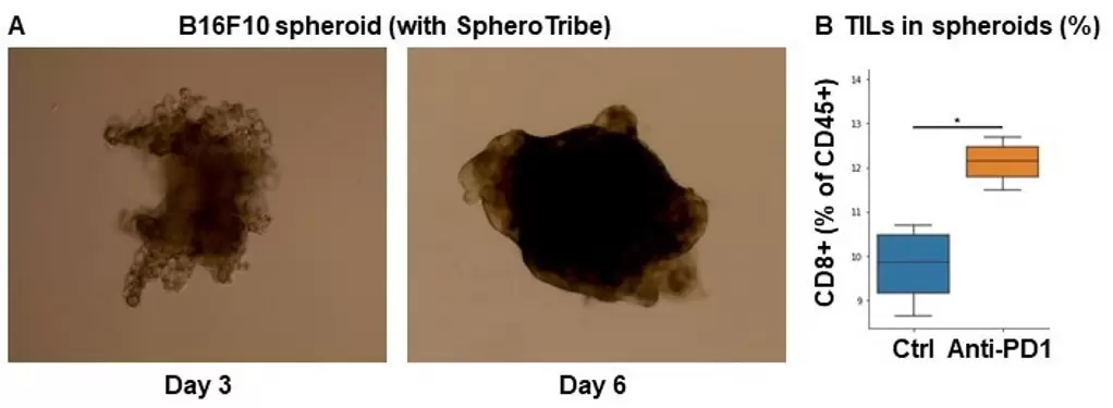
Fig.2. Immune infiltration of B16F10 spheroids after immune checkpoint blockade.
A. 10,000 B16F10 cells were grown for 6 days as spheroids using SpheroTribe.
B. 100,000 PBMC from murine spleen were activated with IL-15 (40 ng/mL), incubated with anti-PD1 (10 µg/mL) for 1h and added on B16F10 spheroids for 3 days.
Graph shows flow cytometry quantification of differential lymphocyte infiltration after spheroid dissociation according to treatment. N=4. Mann-Whitney U Test, p-value<0.05. [1] https://doi.org/10.3389/fonc.2022.898732. Image credits: Guillaume Mestrallet, PhD - Tisch Cancer Institute, Icahn School of Medicine at Mount Sinai, New York
- Catalog Number
TDA-SPK-MINIKIT-IDY - Supplier
Idylle - Size
- Shipping
RT

