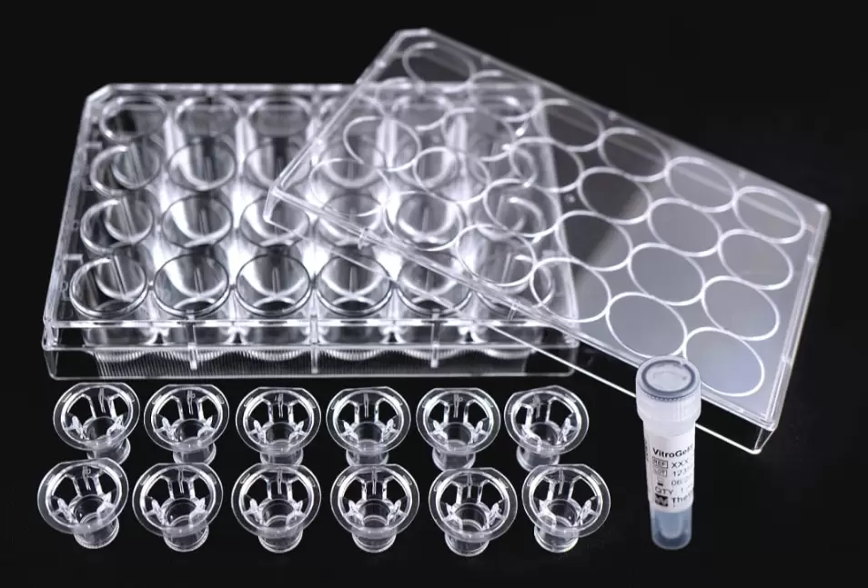
VitroGel® Cell Invasion Assay Kit
Explore how different matrix strengths, ligands, chemokines, growth factors, and more influence cell invasion
Product Description
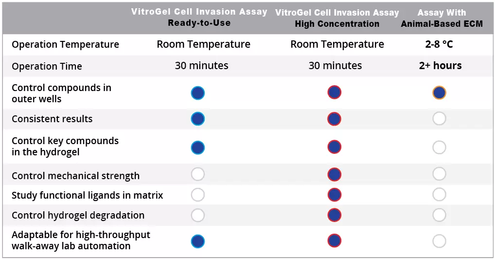
- Full Control of ECM properties – Unlock the understanding of key ECM factors on cell migration/invasion. Gain new knowledge that was not capable with animal-based ECM)
- Synthetic hydrogel with batch-to-batch consistency: Accurate and reproducible results
- Easy to use at room temperature operation: Supports lab automation and high-throughput applications
Cell invasion, a vital process in various biological contexts, can be pivotal in embryonic development, immunosurveillance, and wound healing while also playing a concerning role in cancer metastasis. Traditional in vitro invasion assays have relied on animal-based extracellular matrices, which come with challenges like undefined components, batch-to-batch variability, and cumbersome temperature-sensitive protocols, thus leading to inconsistent and inaccurate studies.
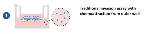
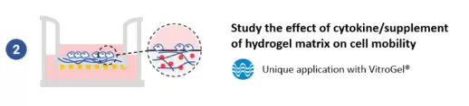
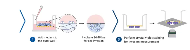
This ready-to-use VitroGel-Based Cell Invasion Assay Kit is powered by VitroGel® hydrogel (versatile, xeno-free, bio-functional hydrogel that closely mimics the physiological extracellular matrix) and coupled with our premium quality VitroPrime™ Cell Culture Inserts, allowing more accurate and consistent invasion and migration studies than animal-based ECM. With the kit, researchers can not only conduct (1) traditional invasion assays by introducing cytokines, chemokines, growth factors, cells, serum, and pharmacological agents to the outer well but also (2) adjust them within the hydrogel matrix to assess cell invasion.
The complete range of kits consists of this ready-to-use VitroGel® Cell Invasion Assay Kit for traditional cell invasion assay studies and different VitroGel “High Concentration” Cell Invasion Assay Kits where the biophysical and biochemical properties are tunable, allowing researchers to explore how different matrix strengths, ligands, chemokines, growth factors, and more influence cell invasion.
How it works
Work confidently at room temperature. No ice bucket here. VitroGel Cell Invasion Assay Kit is ready-to-use. Just mix with your cells. There is no cross-linking agent required. (Click to learn how gelation works)

Watch the Video Protocol.
Comparison to traditional animal-based ECM
Supporting Data
Invasion of U87-MG Glioblastoma cells towards an FBS gradient
- Cell Invasion Assay Kit: VitroGel Cell Invasion Assay Ready-to-use
- Insert: VitroGel Hydrogel Matrix
- Outer well: No serum v.s. 20% FBS
- Cell incubation time: 48 hrs
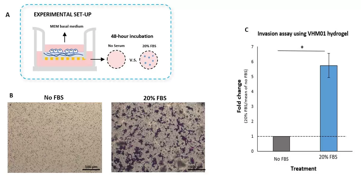
Fig. 1. Invasion of U87-MG glioblastoma cells through VitroGel Hydrogel Matrix caused by a serum gradient.
A. Schematic representation demonstrating the invasion assay cell culture set-up. B. U-87 MG cell invasion was visualized by performing crystal violet staining followed by light microscopy. The images show the membrane inserts from control group (No FBS) and 20% FBS conditions. Images were obtained with a Zeiss microscope at a 10X magnification. C. Fold change of U87-MG cell invasion between control and 20% FBS groups. The control group was normalized to 1. The asterisk (*) stands for p<0.05.
Unique application with VitroGel
Evaluating chemotaxis by adjusting the growth factors compositions within VitroGel Hydrogel Matrix
- Cell Invasion Assay Kit: VitroGel Cell Invasion Assay, Ready-to-use
- Insert: VitroGel Hydrogel Matrix with TGF-β1 v.s. without TGF-β1
- Outer well: No serum v.s. 20% FBS
- Cell Incubation time: 24 hrs
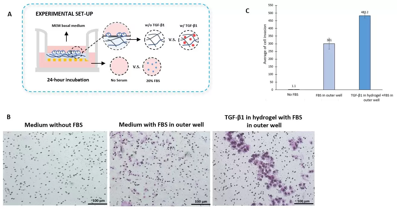
Fig. 2. TGF-β1 inside of VitroGel hydrogel matrix induces invasion of U87-MG glioblastoma cells.
A. Visual representation of invasion assay setup. B. Light microscopy images demonstrating cell invasion in the different groups after crystal violet staining. Images were obtained with a Zeiss microscope at a 10X magnification. C. Mean of U87-MG cell invasion for each of the experimental conditions.
- Catalog Number
IA-VHM01-1P-TW - Supplier
TheWell - Size
- Shipping
RT