Exo-Check Exosome Antibody Array
| Specifications | |
|---|---|
| Product Category: | Exosome Antibody Arrays |
Product Description
This semi-quantitative Exo-Check Exosome Antibody Array provides you with a robust system to check for expression of well-known exosomal markers.
- Requires as little as 50 µg of exosomal protein for detection
- Includes eight antibodies for known exosome markers
- Compatible with most exosome isolation methods, include the ExoQuick® family of reagents and ultracentrifugation
- Includes a control for cellular contamination, a background control (blank spot), and a labeled positive control for HRP detection
- Can be used to evaluate relative abundance of certain exosome markers from a given set of samples
The array has 12 pre-printed spots and features 8 antibodies for known exosome markers (CD63, CD81, ALIX, FLOT1, ICAM1, EpCam, ANXA5 and TSG101), a GM130 cis-Golgi marker to monitor any cellular contamination in your exosome isolations, a positive control spot derived from human serum exosomes, and a blank spot as a background control. The kits come complete with a secondary detection mixture conjugated to HRP.
Supporting data
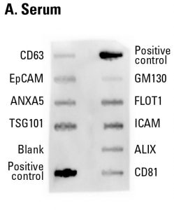
Fig.1. Sample data showing serum-derived exosome detection with an Exo-Check Exosome Antibody Array. The array was exposed to 50 µg of exosome proteins isolated from pooled normal human serum using ExoQuick®. The positive control bands should provide strong signals to indicate that all of the detection reagents are working properly. The various exosome antibody spots will provide varying levels of signal, depending upon the source of the isolated exosomes. The blank band serves as a background control and should not have any signal. The GM130 control will only show signal above background if there is cellular contamination in your exosome preparations.
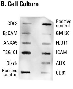
Fig.2. Sample data showing cell culture-derived exosome detection with an Exo-Check Exosome Antibody Array. The array was exposed to 50 µg of exosome proteins isolated from HEK293T cells cultured in SBI’s ExoFBS exosome-depleted media using ExoQuick-TC®. The positive control spots should provide strong signals to indicate that all of the detection reagents are working properly. The various exosome antibody spots will provide varying levels of signal, depending upon the source of the isolated exosomes. The blank band serves as a background control and should not have any signal. The GM130 control will only show signal above background if there is cellular contamination in your exosome preparations.
- Catalog Number
EXORAY200B-4-SBI - Supplier
SBI System Biosciences - Size
- Shipping
Blue Ice
