Exosome-depleted FBS Media Supplement Heat Inactivated - USA Certified
| Specifications | |
|---|---|
| Product Category: | Cell Culture Supplements |
| Cell Type: | Other |
Product Description
Exo-FBS™ Exosome-depleted FBS
- Exosome-sized and CD63 vesicles removed
- No detectable bovine exosomes by Western blot
- No detectable bovine microRNAs by qPCR
- Same cellular growth rates supported as standard FBS
- Growth rate superior to exosome-depleted FBS from other suppliers
- Use the same as standard FBS (add 10% in DMEM or RPMI)
- Optimized for exosome research with numerous peer-reviewed publications
Exosomes are 60-120 nm membrane vesicles secreted by most cell types in vivo and in vitro. Exosomes are found in blood, urine, amniotic fluid, malignant ascite fluids, urine and in media from cells in culture. Exosomes contain distinct subsets of microRNAs depending upon the cell type from which they are secreted. Standard growth medium for most cells in culture require fetal bovine serum (FBS) as a growth supplement to DMEM. FBS is derived from bovine (cow) serum and contains a high abundance of cow exosome vesicles. These exosomes can interfere or cause significant background issues when studying the exosomes secreted from your cells of interest in standard culture conditions.
SBI has developed an exosome-depleted FBS growth supplement called Exo-FBS that has been stripped of bovine exosomes. Exo-FBS supports equivalent growth of many types of cells in culture, contains very low levels of cow CD63 positive exosomes and does not have any measurable bovine microRNAs. Perform your studies of cellular secreted exosomes in culture without the worry of contaminating cow exosomes in your experiments. No ultracentrifugation required. Available as standard FBS supplement or heat inactivated FBS media supplement (treated at 65°C for 15 minutes before bovine exosome removal) in two sizes, each.
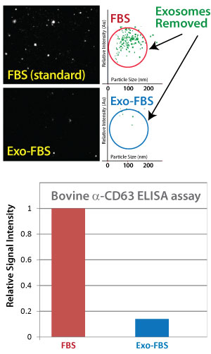
Exosomes removed in Exo-FBS
 NanoSight particle analysis of FBS and Exo-FBS
Standard FBS and Exo-FBS samples were diluted 1:1000 and then analyzed for particle size and abundance using a NanoSight LM10 instrument. The standard FBS sample shows a significant amount of exosome-sized microvesicles where the Exo-FBS exosome-depleted FBS sample has a drastic reduction in exosome particles.
Exo-FBS is depleted of bovine CD63 exosomes
The tetraspanin CD63 protein is a common marker for exosomes. We utilized a bovine-specific anti-CD63 antibody to develop an Enzyme Linked Immunosorbent Assay (ELISA). Equal volumes (50 ul) of either standard FBS or Exo-FBS depleted media supplement were used in this ELISA assay. Amounts of CD63-positive bovine exosomes dramatically reduced. The results are graphed and normalized to the signal level of standard FBS.
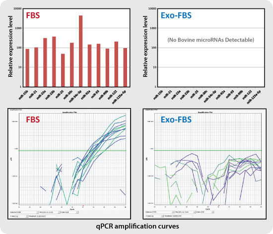
Bovine microRNAs are absent in Exo-FBS

Standard FBS and Exo-FBS media supplements (4 ml) were treated with Trizol extraction methods to recover exosome RNAs. RNA was converted to cDNA and 72 individual bovine microRNAs were measured by qPCR using SBI's QuantiMir system. Of the 72 microRNAs tested, 12 yielded amplification curves in the FBS sample but not in the Exo-FBS sample.
Bovine microRNAs present in FBS are no longer detectable in Exo-FBS - No more cow microRNAs!
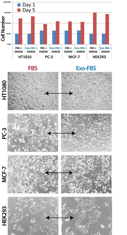
Exo-FBS media supports robust growth of cells in culture equal to standard FBS media

Complete medium either with 10% standard FBS or with 10% Exo-FBS supplement. HT1080 fibrosarcoma cells, PC-3 prostate cancer cells, MCF-7 breast cancer cells and HEK293 cells were seeded at either 10,000 or 20,000 cells and then cultured under standard conditions at 37°C with 5% CO2 for 5 days in the medium indicated. The cells were imaged for growth numbers and morphologies. Equivalent growth and morphologies were observed for standard FBS and Exo-FBS media tested across these 4 cell lines tested.

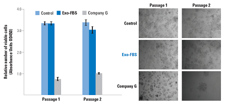
If you think one company's exosome-depleted FBS is just like another's, take a look at the data above. Exo-FBS clearly supports better growth than Company G's exosome-depleted FBS. Whether you look at cell viability or cell morphology, Exo-FBS is the obvious reagent of choice for robust cell growth for extracellular vesicle studies.

