AgarSqueezer Device (2 devices + 1 microscope holder)
Confine and study your cell behavior within a physiological rigidity range

Product Description
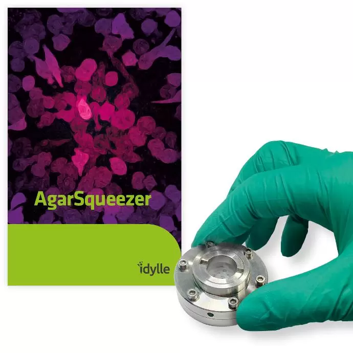
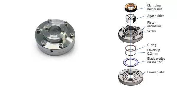
AgarSqueezer is a microscope slide chamber equipped with a molded agar-based compression system. Use it to apply an instant & homogeneous compression on your cells, and study their response to short and long-term confinement.

Compatible Assays
- Live imaging*: cell viability, proliferation, migration, morphology, nuclear deformability, cytoskeleton reorganization, cell tracking, etc
- Standard molecular analysis (immunostaining, western blot, qPCR, flow cytometry, etc): changes in gene/protein expression, proliferation, morphological analyses, etc.
*Cells can be added with fluorescent probes or stimulated with drugs during confinement.
Kit Content
2 AgarSqueezer Devices, 16G and 20G flat-cut needles, 1 autoclavable screwdriver and (optional) 1 sample holder for use with live cell imaging chambers. The kit is also available in a small version with 1 device.
Silicium Wafers
In addition to the basic kit, we recommend using a silicium wafer to mold your confining pillars in agarose. We provide four different wafers to mold pillars of various heights. The wafers have to be purchased separately, choose among our four pillar heights available (2,5μm, 5μm, 30μm, 100μm) depending on your desired application.

Fig.2. Different compression heights are available for confinement.
How To Use
Applications
AgarSqueezer has been successfully used to confine adherent and non-adherent cells, including:
- Human: primary T-lymphocytes, TF1 & ML2 leukemic cells, HS27A fibroblasts, MCF10A breast cells, MDA-MB-231 breast cancer cells, U-2 OS osteosarcoma cells, PC-3 & DU 145 prostate cancer cells, HT29 & HCT116 colorectal adenocarcinoma cells and HT1080 fibrosarcoma cells
- Murine: megakaryocytes, osteocyte-like cells MLO-Y4, primary muscle cells & primary dendritic cells
- Plant: cells from Arabidopsis roots
- 3D cell cultures: mice gastruloïds
Imaging Modes
Inverted microscopes with any imaging mode: phase-contrast, epifluorescence, confocal, etc.
Supporting Data

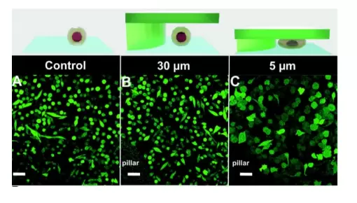
Fig.3. Compression of immature TF1-GFP hematopoietic cells Quantification of cell morphology under confinement. (A–C): Morphology of immature TF1-GFP hematopoietic cells for control (A) and for 30 μm and 5 μm (B and C, respectively). Scale bar = 20 μm.
From A. Prunet et al. Lab on Chip, 2020.
- Catalog Number
CRI-SOF-2-IDY - Supplier
Idylle - Size
- Shipping
RT

