NTA-based Exosome Characterization Services
Nanoparticle Tracking Analysis-based Exosome Characterization Services
Get quality data on exosome particle size and concentration
- Understand the quality of your exosome preps
- Leverage SBI’s long-term experience working with exosomes
- Send us your biofluid or exosome prep and receive data in about 3 weeks
Get a clearer picture of the quality of your exosomes with SBI's Nanoarticle Analysis Service. Send us your biofluid or prepared exosomes, and we'll send back a report with the particle analysis data which includes the mean and mode diameter size as well as particle concentration. Fast and efficient—turn-around time is approximately 3 weeks—SBI's NTA Analysis Service is an excellent way to characterize your exosome preps, with no need for your own NTA instrument.
What’s NTA?
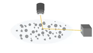
SBI uses NTA measurements that rely on light scattering to extract particle size and the number of particles in a sample (Figure 1A). Briefly, a laser is directed at the sample and the particles in the sample scatter the light, which is imaged by a camera attached to a microscope and operating at 30 frames per second. The NTA software collects data on multiple particles simultaneously, and calculates the hydrodynamic diameter of each particle using the Stokes-Einstein equation.
A. Conventional NTA
All particles are visible to NTA
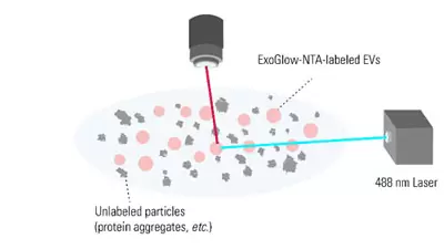
B. Fluorescent NTA
Only ExoGlow-NTA-labeled particles are visible to NTA
Figure 1. The ExoGlow-NTA dye only binds to membranes of intact EVs. Unlike conventional NTA (A), which collects data on all particles in a solution based on light scattering, the fluorescence mode of the NTA instrument selectively detects only the fluorescently-labeled particles—in this case, the ExoGlow-NTA-labeled EVs (B)—and only the data from fluorescently-labeled particles is reported.
What is fluorescent NTA and how is it different from conventional NTA?
Making a great exosome research tool even better, SBI has developed ExoGlow-NTA, a proprietary dye for fluorescent NTA* that enables data collection for only the extracellular vesicles present in a heterogenous sample. The result is more accurate EV/exosomal NanoSight data that ignores protein aggregates, membrane fractions, and other background particles since they do not bind the ExoGlow-NTA dye (Figure 1B) thus providing EV-specific particle size and concentration.