Cell Tracking by Molecular Imaging
Cell Tracking by Fluorescence, Bioluminescence or Positron Emission Tomography (PET)
Cell visualization in vivo and ex vivo for tracking cell fate
Molecular imaging techniques to visualize cell kinetics in small animals have resulted in an explosion in the knowledge of tumors, infectious diseases, and stem cell biology. The sensitivity and accuracy of in vitro and in vivo cell monitoring offers several advantages over traditional methods through animal sacrificing and histological analysis. Molecular imaging, for example, is normally non-invasive and allows for quantitatively assessing tumor growth and the effects of therapy over time.
Easily label cell lines for tracking cell fate after implantation in an animal model or other imaging applications with SBI’s line of bioluminescent, fluorescent, and PET imaging vectors (BLIV) or Lenti-Labeler constructs. Available in a variety of promoter-reporter combinations that enable multiple imaging modalities - fluorescence with copGFP or RFP, bioluminescence with luciferase, and positron emission tomography (PET) with thymidine kinase (TK) - you can select from lentivector (plasmid DNA or lentiviral particles) or minicircle technologies for both integrated and episomal expression.
Choose a lentivector when:
You’re working with difficult-to-transfect cells
You’d like to create stable reporter cell lines
You’d like to integrate your reporter into the genome
Choose a minicircle vector* when you’re working with transfectable cells and:
You’d like episomal expression
You’d like to avoid introducing foreign sequences
Choose your promoter using the following table:
![]()
*Note that Minicircle Parental Vectors are only available to academic/non-profit customers. Researchers at commercial organization may only purchase the pre-made minicircle BLIVs.
Applications
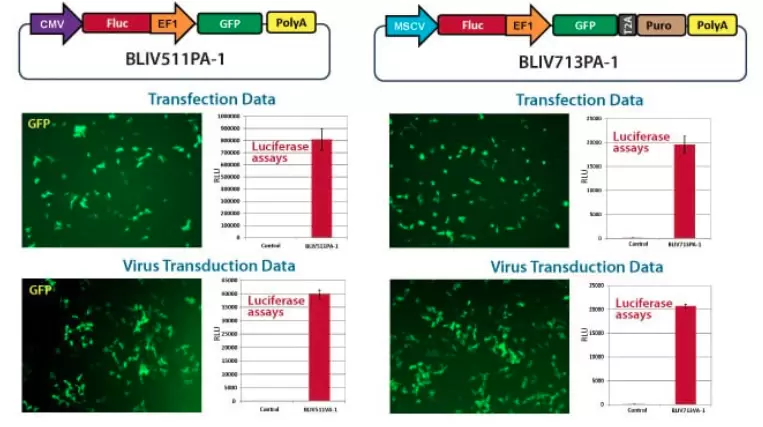
BLIV lentivectors deliver strong luciferase activity after both transduction and transfection

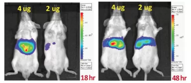
BLIV minicircle vectors deliver strong luciferase activity in small animal models

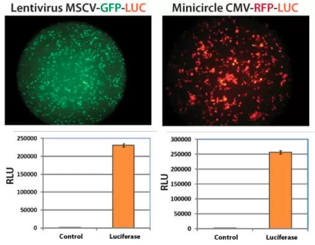
BLIV lentivectors and minicircle vectors can be used to generate stable cell lines with strong luciferase activity
