Protein Arrays
Profile antibodies in bodyfluids or efficiently explore protein-protein and protein-ligand interactions
RayBio Protein Arrays
RayBio's Protein arrays use a 75 mm x 25 mm glass slides spotted with either 8 or 16 identical protein arrays (also called “subarrays”). Within each subarray, the purified protein autoimmunogens along with controls are spotted in triplicate. The slide comes with a 16-well removable gasket and chamber assembly that allows for simultaneous processing of up to 16 samples per slide (8 if using the 8 sample kit).
To achieve even higher throughput, 4 slides can be nested into a tray which matches a standard microplate, allowing for automated processing of 64 arrays (If using the 16 sample kit) simultaneously. The latest automated liquid handling work stations can accommodate several trays at once, allowing hundreds of samples to be processed per day.
Features
- Less sample, more data: only very small sample volumes are needed quantification of up to 48 targets
- Les than 8-hour processing time
- No interference between immobilized proteins (unlike bead-based multiplex assays)
- No dedicated equipment required; Array scanning and Analyses service available
- Customizable – create a custom array from our list of available targets
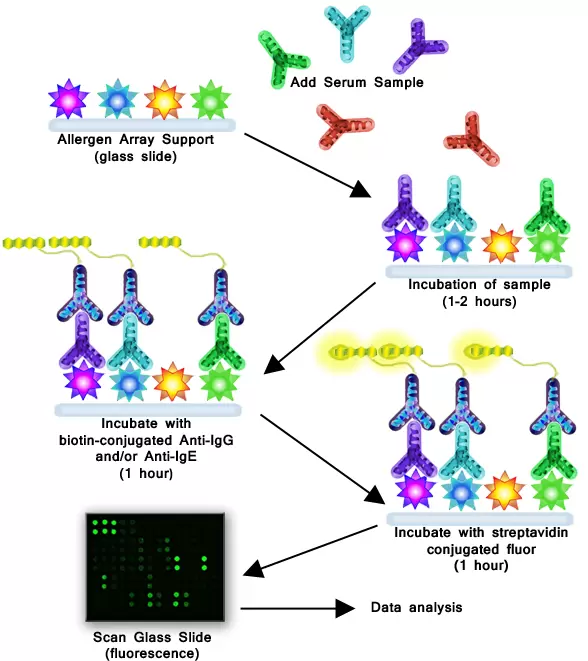
How It Works
Allergen Protein Array as example: A panel of allergen proteins is printed in multiple identical arrays on a standard slide. After a blocking step, samples are incubated with the arrays. Nonspecific antibodies are then washed off, and the arrays are incubated with Biotin-conjugated anti-human IgG and/or IgE detection antibodies, followed by a streptavidin-conjugated fluor. Signals are then visualized using a fluorescence laser scanner.
Allergen Protein Arrays
The RayBio® Allergen Protein Arrays are semi-quantitative multiplex protein arrays developed for the detection of antibodies to common allergens. Recombinant allergen proteins or extracted native allergen proteins are first spotted onto a solid glass slide surface, and then probed with serum to measure the presence and levels of serum-specific IgE and/or IgG antibodies targeting these allergens.
Cancer Autoantibody Arrays
The RayBio® Cancer Autoantibody Arrays are semi-quantitative multiplex protein arrays developed for the detection of cancer-related auto-antibodies. Recombinant proteins previously associated with cancer are spotted onto a solid glass slide surface, and then probed for the presence of serum-specific antibodies to these targets. These detection kits can then be applied in screening specific autoantibodies based on IgA or IgG levels present in patient serum samples.
Human Autoimmune Disease IgG Autoantibody
RayBio® Autoimmune Disease (AID) IgG Autoantibody Array contains 33 human proteins and double strand DNA(dsDNA) for simultaneous detection of human IgG autoantibodies presented in blood serum samples of common 10 autoimmune diseases, such as systemic lupus erythematosus (SLE), Sjøgren's syndrome, Rheumatoid arthritis, primary biliary cirrhosis, etc.
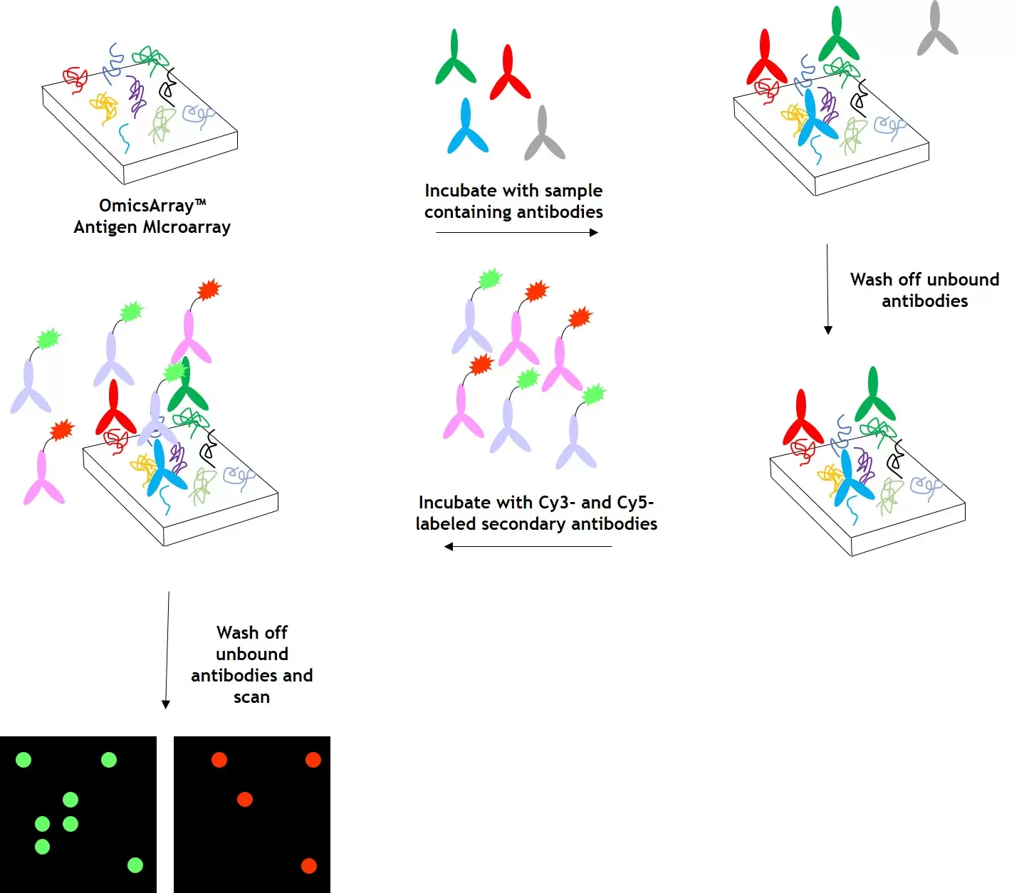
OmicsArray™ Antigen Microarrays
The OmicsArray™ Antigen Microarrays developed by GeneCopoeia are high-throughput protein microarrays for many applications, such as autoantibody profiling, biomarker detection, pathogen-specific antibody identification, and more. Each array contains superior-quality purified antigens printed onto nitrocellulose filters, which are adhered to glass slides. Antigens known to be associated with specific diseases or pathogens are chosen based on a thorough review of peer-reviewed publications. OmicsArray™ Antigen Microarrays, including a large number of whole-protein antigens specifically focused on autoimmune disease research, offer the best combination of number of antigens per array (up to 120) with number of samples that can be processed per slide (up to 15).
How it works
Arrays are incubated with patient samples, and any autoantibodies in the samples bind to their cognate antigens on the array. The arrays are washed to remove unbound autoantibodies and other proteins, then co-incubated with Cy3- and Cy5-labeled secondary antibodies. The dual labeling strategy is intended to distinguish between immunoglobulin (Ig) subtypes present within samples. After washing to remove unbound secondary antibodies, signals are detected using a microarray scanner (e.g., GenePix® 4000B, InnoScan 710, or equivalents). The raw data is then be analyzed using GenePix® Pro 7.0, or Mapix software.